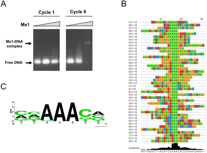
Fig 3. The specificity of DNA binding.
A: Gel shift assays with DNA pools from stages 1 (first) and 8 (last) of the SELEX procedure with Ms1 270–375. Positions of free and bound DNA are indicated. B: Sequencing results from the final SELEX pool after stage 8. Only random region shown, constant region is omitted. C: Web logo [40] plot of the consensus DNA recognition motif of ABD2.

