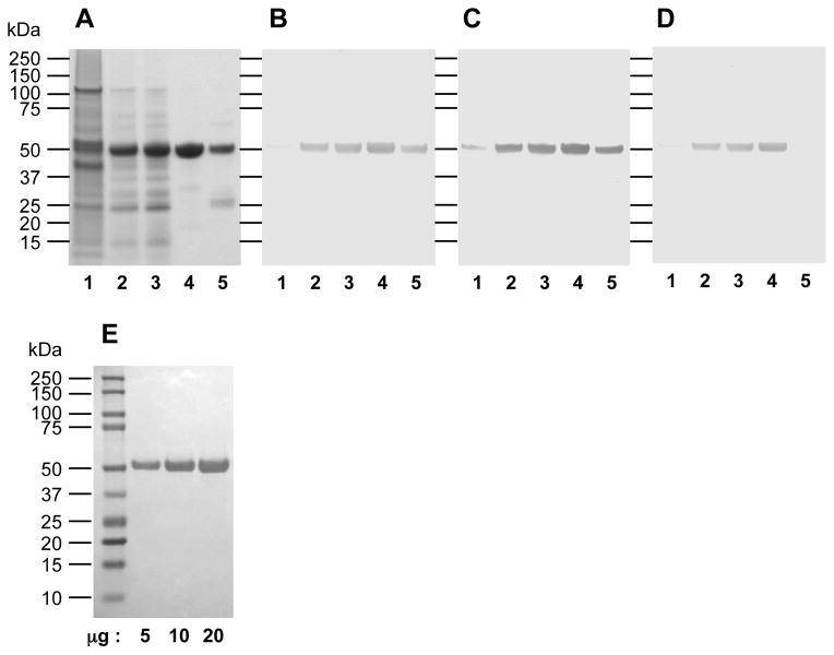
Figure 2.
SDS-PAGE and Western-blot analysis of RgpB-6His purification steps as described in Table 1. Lanes: 1, initial culture supernatant; 2, after acetone precipitation; 3, after ion exchange chromatography on DE-52 matrix; 4, eluted fraction from Ni-Sepharose affinity chromatography; 5, flow-through fraction from Ni-Sepharose chromatography. 10 μg of each sample were electrophoresed on a 4–12% BisTris SDS-PAGE gel and stained with Simple Blue Safe Stain (Invitrogen) (A). Alternatively, 1 μg of proteins were electrophoresed under the same condition and electro-blotted onto nitrocellulose membrane. After blocking with skim milk, membranes were probed with rabbit pAb antiRgp (B), or mouse mAb anti-6His detecting exclusively hexa-histidine tags (Roche, Indianapolis, IN, USA) (C).

