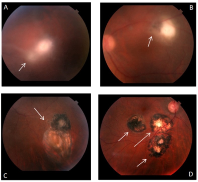
Fig 1. Colour retinography showing the various stages of eye lesions caused by Toxoplasma gondii infection in Brazilian patients.

In (A) the arrow indicates the region with an acute exudative chorioretinal lesion ("lighthouse in the fog") and cloudy vitreous. In (B) the arrow indicates a chorioretinal lesions in the healing process—the patient had good clinical response to treatment and scar edges in definition. In (C) the arrow indicates presentation of an old chorioretinal scar and an old chorioretinal satellite lesion with pigment mobilization. In (D) chorioretinal scaring with well-defined edges indicated by the arrows with visualization of the sclera.
