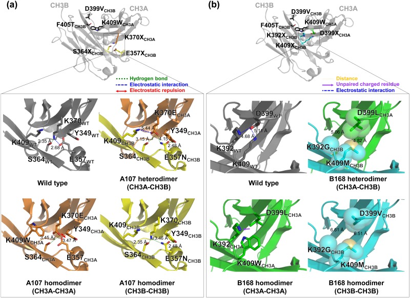
Fig 4. Modeled CH3 domain interface structures of the A107 (a) and B168 (b) variants based on the crystal structure of EW-RVT variant (PDB code 4X98).
Each upper panel shows the CH3A-CH3B heterodimer structure of the W-VT variant to highlight the targeted mutation sites in LibA1 (a) and LibB1 (b) (as highlighted in the square) on the opposite side of the parent W-VT variant pair (K409WCH3A-D399V/F405TCH3B) at the CH3 interface. The lower panels shows a close-up view of the newly introduced mutation pair in A107 (K370ECH3A-E357NCH3B) (a) and B168 (D399LCH3A-K392G/K409MCH3B) (b) in a CH3A-CH3B heterodimer, CH3A-CH3A homodimer, and CH3B-CH3B homodimer, compared to the wild type CH3 homodimer. Details are described in the text.

