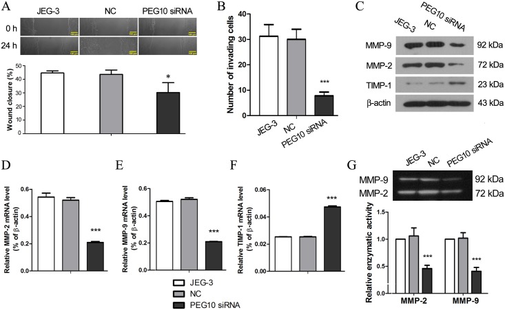
Fig 3. PEG10 silencing inhibited migration and invasion of JEG-3 cells.
(A) Migration of PEG10-silenced JEG-3 cells was evaluated by in vitro scratch wound assay. (B) Matrigel Transwell assay was performed to assess invasiveness of PEG10-silenced JEG-3 cells. Expression of the key players of cell invasion including MMP-2, MMP-9 and TIMP-1 was examined by (C) Western blotting and (D-F) real-time PCR. (G) Gelatin zymography was performed to determine the enzymatic activities of MMP-2 and MMP-9 which were from the conditioned culture medium. The figure shows the representative images of three independent experiments and data are expressed as the mean ± SD. Significance versus non-transduced JEG-3 cells, *p< 0.05, ***p< 0.001.

