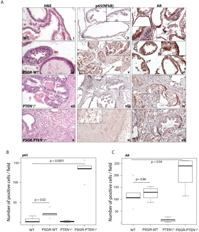
Figure 4. Androgen receptor (AR) expression pattern is correlated with p65 (NF-κB) expression pattern.
A. Staining shows expression pattern of p65 (NF-κB) and AR in prostate sections of 12 month old mice. PSGR-WT mice show both cytoplasmic and nuclear localization of p65 and AR (v and vi). Nuclear localization is correlated with protein activity. PSGR-PTENΔ/Δ mice show increased levels of nuclear p65 and AR in the stromal compartment of the prostate (xi and xii). B. Box plots represent the average number of positive cells per field for each genotype. Only nuclear p65 and AR staining was considered as positive (n=4mice/group), Comparison of p65 staining between WT and PSGR over-expressing mice is significantly increased (p-value = 0.02), whereas concomitant deletion of PTEN and PSGR over-expression further increases p65 nuclear staining (p-value = 0.0001). C. For AR, there is no significant difference in stromal AR staining (p-value = 0.86) between WT and PSGR over-expressing mice, and deletion of PTEN alone is enough to induce a significant loss of stromal AR (p-value = 0.009). Double mutant mice, however, show a statistically significant increase in stromal AR staining (p-value = 0.04), suggesting deletion of PTEN is necessary for PSGR to induce expression of stromal AR.

