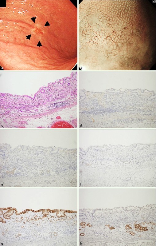
Fig. 2.

Undifferentiated early adenocarcinoma with gastric phenotype as one of the representative WOS-negative gastric neoplasias. a A slightly reddish and whitish colored 0-IIc type neoplasia (arrow) was observed at the lower body of the stomach with white light endoscopy. b WOS was not detected by M-NBI. c Hematoxylin and eosin staining of the resected specimen shows signet ring cell carcinoma cells infiltrating the intramucosal layer. d No adipophilin postive cells were observed. e Neoplastic cells and the adjacent non-neoplastic epithelium were negative for CD10. f Neoplastic cells and the adjacent non-neoplastic epithelium were negative for MUC2. g Neoplastic cells located on the surface and the residual non-neoplastic epithelium were positive for MUC5AC. h Neoplastic cells were negative for MUC6, but the non-neoplastic epithelium at the deep portion was positive for MUC6 focally.
