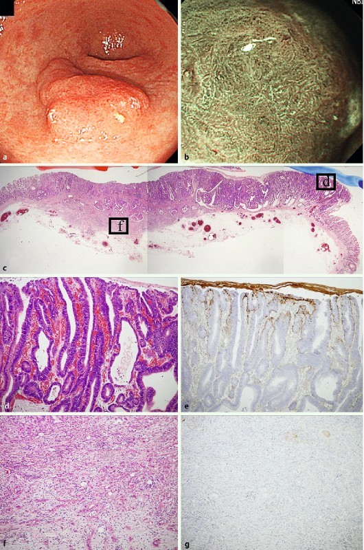
Fig. 3.

Mixed type (mixed predominatly differentiated type) early adenocarcinoma of gastrointestinal phenotype as a representative WOS-positive gastric neoplasia. a. A slightly elevated reddish colored 0-IIa type neoplasia was observed at the gastric antrum with white light endoscopy. b M-NBI findings showed the irregular WOS at the oral side of the tumor. c Hematoxylin and eosin staining of the resected specimen. The tumor was composed of an intramucosal well to moderately differentiated tubular adenocarcinoma component and an invasive poorly differentiated adenocarcinoma component visualized at low magnification. d High maginification of box d in Fig. 3 c showed that the tumor glands were composed of well differentiated tubular adenocarcinoma. In this area, WOS was detected by M-NBI. e Positive adipophilin expression was only observed in the well differentiated tubular adenocarcinoma component. f High maginification of box f in Fig. 3 c shows poorly differentiated adenocarcinoma cells invading the submucosal layer. g Adipohilin expression was not detected in the poorly differentiated adenocarcinoma component.
