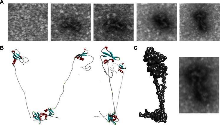
Fig. 3.
Electron microscopy validates the shape of the HP1γ homodimer. a Electron microscopy (EM) images of purified HP1γ. b Homology-based model of the HP1γ homodimer. c Superimposition of the homodimer structure shows that the predicted model fits nicely with the shape determined by EM imaging. Similar observations have been recently obtained for the yeast HP1 proteins, SWI6 [9]

