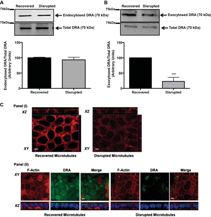
Fig. 5.
Role of microtubules in basal DRA endocytosis and exocytosis. A: Caco-2 monolayers were incubated with sulfo-NHS-SS-biotin, and microtubules were disrupted and incubated at 4°C. Microtubule recovery upon return to 37°C was prevented by addition of nocodazole to the medium or was allowed to proceed in the absence of nocodazole. Total protein lysates were incubated with NeutrAvidin-agarose beads to pull down biotinylated proteins. Total and biotinylated fractions were run on a 7.5% SDS-polyacrylamide gel and then immunoblotted with anti-DRA antibody. DRA bands in the representative blot were quantified by densitometric analysis. Results are shown as ratio of endocytosed to total DRA. Values are means ± SE of results from 3 different blots. B: cell surface Caco-2 cells were masked with sulfo-NHS-acetate. Cells were then incubated at 4°C to disrupt microtubules and allowed to recover, or not, in the absence or presence of nocodazole. Amount of newly exocytosed protein was assessed by cell surface biotinylation. Immunoblot was probed with anti-DRA antibody and quantitated by densitometric analysis. Results are shown as ratio of endocytosed to total DRA. Values are means ± SE of results from 3 different blots. ***P < 0.001 vs. recovered. C: Caco-2 cells were subjected to cold exposure + nocodazole treatment to induce microtubule disruption. I: cells were stained for α-tubulin (red) to confirm microtubule disruption and recovery. II: Caco-2 cells were immunostained for localization of DRA (green) and Alexa Fluor 568-conjugated phalloidin (actin; red), and DAPI was used to label nuclei (blue) in cells with recovered and disrupted microtubules.

