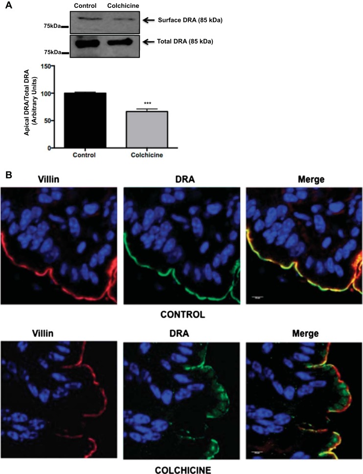
Fig. 6.
Disruption of microtubules in vivo reduces DRA surface expression. A: ex vivo biotinylation was performed in colons from control and colchicine-treated (3 mg/kg) mice 24 h after administration of the drug. Total lysates from control and treated mice were incubated with NeutrAvidin-agarose beads overnight at 4°C to pull down biotinylated proteins. Biotinylated and total fractions were run on a 7.5% gel and then transferred to a nitrocellulose membrane. Membrane was probed with anti-DRA antibody, and protein bands were quantified by densitometric analysis. Results are shown as ratio of surface (apical) to total DRA. Values are means ± SE of results from 3 different experiments. ***P < 0.006 vs. control. B: immunofluorescent staining [DRA (green) and villin (red)] in colonic tissue sections from control and colchicine-treated mice. Nuclei were stained with SlowFade with DAPI (blue). A representative image from 3 different animals for each treatment is shown. Images were obtained using a confocal microscope (LSM 710, Zeiss) with a ×63 objective. Control and colchicine images were obtained in the same plane.

