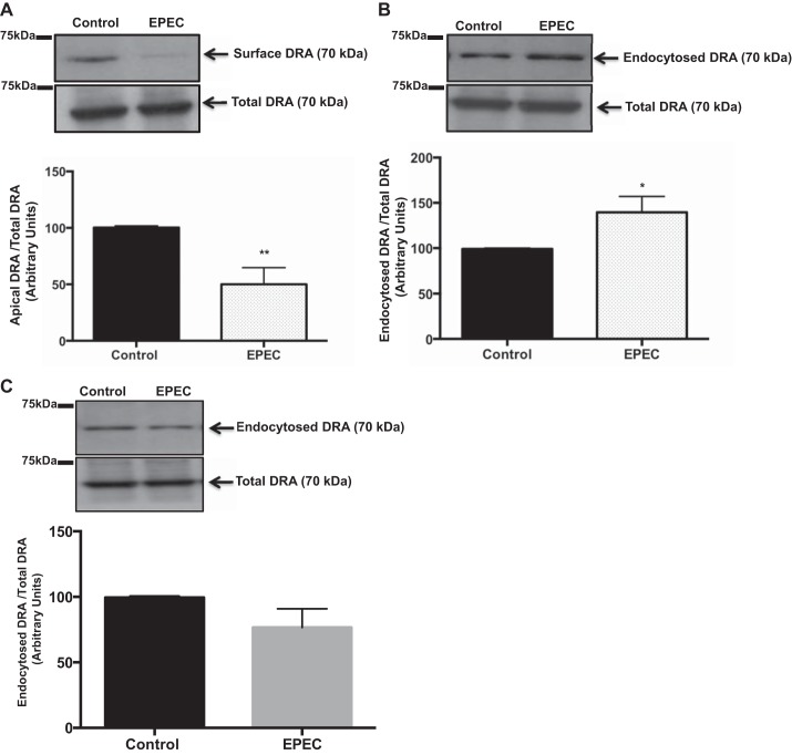
Fig. 7.
Enteropathogenic Escherichia coli (EPEC) decreases DRA surface expression partly via increased DRA endocytosis in Caco-2 cells. A–C: cells infected with EPEC for 60 min were subjected to cell surface biotinylation (A), reverse cell surface biotinylation (B), or reverse biotinylation on ice (negative control, n = 5; C). NeutrAvidin-agarose beads were used to pull down biotinylated proteins. Biotinylated and total protein fractions were run on a 7.5% SDS-polyacrylamide gel. Blot was immunostained with rabbit anti-DRA antibody. Biotinylated fractions (A and B) represent apical and endocytosed pool of DRA, respectively. Densitometric analysis presented below each blot is representative of 3 separate experiments. Results are shown as ratio of apical to total DRA (A) and endocytosed to total DRA (B and C). Values are means ± SE of 3 different experiments. *P < 0.05 and **P < 0.004 vs. control.

