Fig. 10.
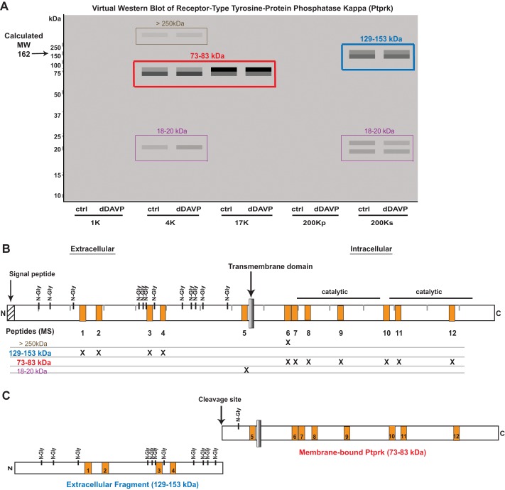
In mpkCCD cells, receptor-type tyrosine-protein phosphatase-κ (Ptprk) is cleaved into 2 fragments. A: virtual Western blot of Ptprk. Colored boxes indicate distinct bands that contain MS-identified peptides. B: map of Ptprk protein showing locations of mass spectrometry-identified peptides as orange boxes (1–12). MW regions in the gel in which these peptides were found are indicated by “X.” C: deduced cleavage products of Ptprk and the cleavage site identified by Jiang et al. (11). N-Gly, N-glycosylated.

