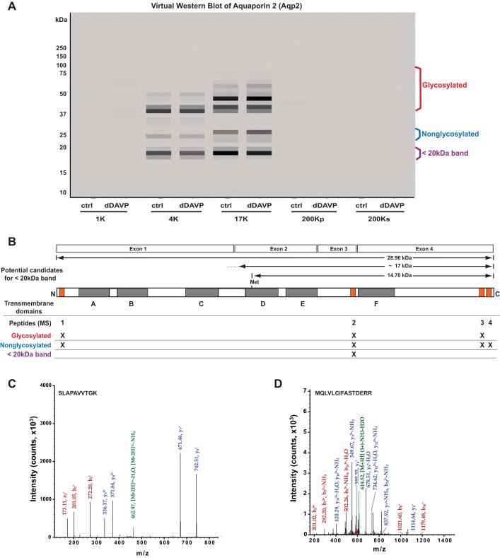
Fig. 12.
In mpkCCD cells, AQP2 (Aqp2) peptides map to 3 broad bands. A: in a virtual Western blot of AQP2, peptides map to bands known to be associated with the nonglycosylated and glycosylated forms (labeled in blue and red, respectively). An additional band was identified at <20 kDa (purple label). B: AQP2 structure showing transmembrane domains (shaded, A–F) and the corresponding 4 exons that code for the full-length protein. Orange boxes indicate the map of 4 mass spectrometry-identified peptides to the Aqp2 protein. MW regions in the gel in which these peptides were found are indicated by “X.” C: one of the spectra for peptide 2 (SLAPAVVTGK). Green, precursor peak; blue, y-series peaks; red, b-series peaks. D: one of the spectra corresponding to a new peptide (MQLVLCIFASTDERR), identified multiple times by targeted search of original mass spectrometry data. Green, precursor peak; blue, y-series peaks; red, b-series peaks. This peptide corresponds to the NH2-terminal peptide in hypothetical AQP2 protein coded by exons 2, 3, and 4. It was found only in the gel slice corresponding to 18.5 kDa.

