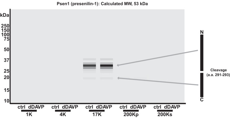
Fig. 9.
In mpkCCD cells, presenilin (Psen1) is cleaved into 2 fragments of 32 and 19 kDa, residing in the 17-kDa fraction. In the virtual Western blot, Psen1, a 53-kDa protein, appears as 2 bands of 32 and 19 kDa. NH2- and COOH-terminal fragments identified on the basis of mapping mass spectrometry-identified peptides are indicated with arrows. A cleavage site, between amino acids 291 and 293 (UniProt no. P49769), is shown at right.

