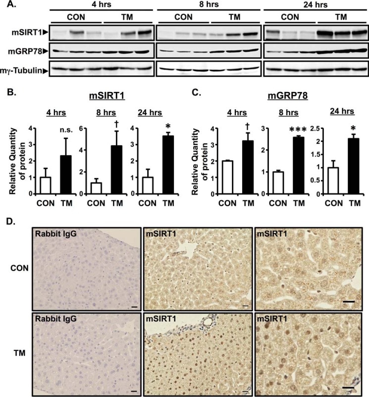
FIGURE 2.
ER stress increases the protein expression of SIRT1 in vivo. A–C, C57BL/6J mice were intraperitoneally injected with TM (1 μg/g of body weight), and liver tissues were harvested at 4, 8, or 24 h after injection. Expression of mSIRT1 and mGRP78 protein was determined by Western blotting analysis of whole cell lysate. Mouse γ-tubulin (mγ-tubulin) was used as a loading control. The relative quantity of mSIRT1 (B) and mGRP78 (C) protein was analyzed by Image Gauge software. Values are the mean ± S.E. (error bars) (n = 3 for B and C). *, p < 0.05 versus control; ***, p < 0.001 versus control; †, p < 0.1 versus control as assessed by Student's t test. n.s., not significant. D, immunohistochemical analysis of SIRT1 in TM-injected mouse liver tissue. Livers were collected 24 h after intraperitoneal injection with TM. Rabbit IgG was used as a negative control. Scale bars indicate 20 μm. CON, control.

