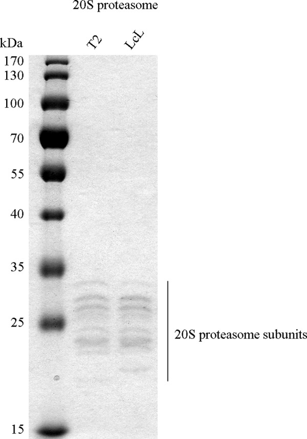
FIGURE 8.

Purity of 20S proteasome preparation. 20S proteasomes purified from T2 and lymphoblastoid (LcL) cell lines (1 μg) were separated by SDS-PAGE and stained by Coomassie Blue to detect possible contaminations. Only the characteristic bands of 20S proteasome, with sizes between 21 and 31 kDa, are visible, confirming the high purity of the proteasome preparation. For instance, no contamination with Hsp molecules at high molecular mass (above 60 kDa), which are often present in less pure proteasome preparations, could be detected.
