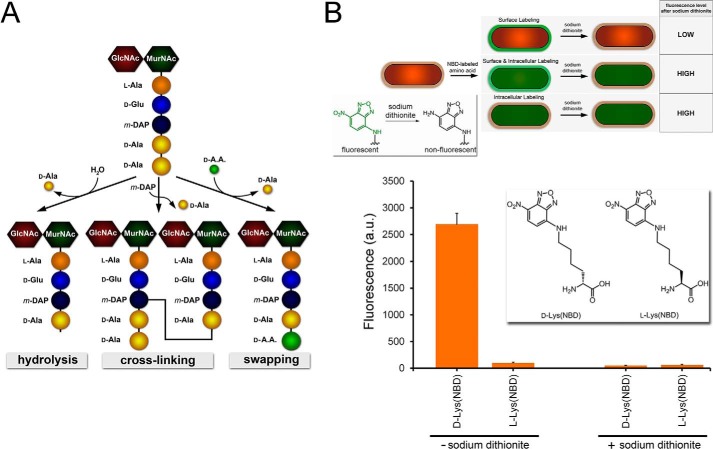
FIGURE 1.
A, schematic showing the monomeric unit of peptidoglycan. The pentapeptide is processed by PBPs CP and TP to generate three possible products, all resulting from the acyl intermediate following the release of the terminal d-Ala. B, schematic demonstrating the three possible labeling locations within the bacterial cell. Only extracellular labeling would lead to complete reduction of the signal in the presence of the reducing agent sodium dithionite. Inset, reduction of the nitro group on NBD by sodium dithionite quenches the green fluorescence. B. subtilis cells were labeled with either d-Lys(NBD) or l-Lys(NBD) overnight. Cells were analyzed by flow cytometry. Data are represented as mean ± S.D. (n = 3).

