Fig. 3.
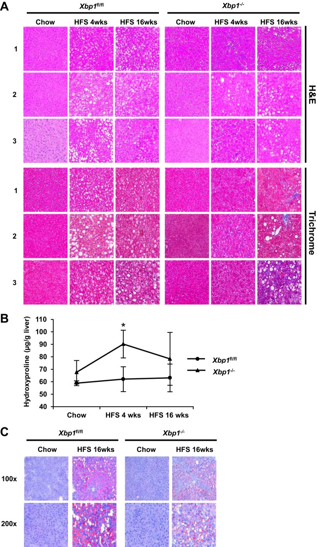
Xbp1−/− mice fed a HFS diet have increased fibrosis. Xbp1−/− and Xbp1fl/fl mice were fed either with chow or a HFS diet for 4 or 16 wk. A: hematoxylin and eosin (H&E; top) and Masson's trichrome staining (bottom) of liver sections (magnification: ×200) from each group were shown. Numbers on the left indicate liver sections from 3 different mice. B: liver hydroxyproline levels were measured. *P = 0.05 compared with HFS-fed Xbp1fl/fl mice. C: representative Oil Red O staining were shown from Xbp1−/− and Xbp1fl/fl mice fed with either chow or a HFS diet for 16 wk. Magnification: top, ×100; bottom, ×200.

