Fig. 2.
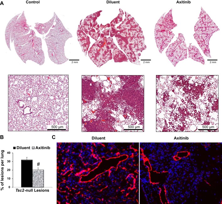
Axitinib treatment inhibits Tsc2-null lung lesion growth and abnormal lymphangiogenesis. A: representative micrographs of hematoxylin and eosin staining of lung sections from control mice and mice with Tsc2-null lesions treated with diluent or axitinib. B: statistical analysis of the percentage of lesions per lung treated with diluent (n = 9) or axitinib (n = 6) was performed as described (13). Values are means ± SE. #P < 0.05 by Student's t-test. C: lung tissue of mice with Tsc2-null lesions treated with diluent or axitinib were analyzed for lymphatic vessels by immunohistochemical analysis with specific anti-lymphatic vessel endothelial hyaluronan receptor 1 antibodies (red). 4′,6-Diamidino-2-phenylindole staining was performed to detect nuclei (blue). Representative images were taken using a Nikon Eclipse TE-2000E microscope (n = 5 per group, magnification: ×20).

