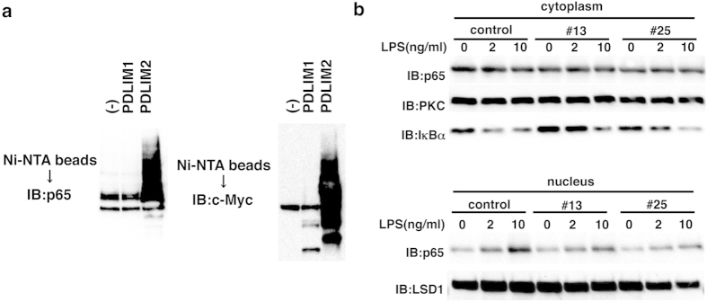
Figure 4. PDLIM1 suppresses LPS-induced nuclear translocation of p65.
(a) Ubiquitination assay for p65 in 293T cells transfected with plasmids encoding histidine-tagged ubiquitin, p65 and PDLIM1, PDLIM2 or empty vector (−). Ubiquitinated proteins were purified with Ni-NTA beads. Polyubiquitination of p65 (left) or autoubiquitination of PDLIM1/PDLIM2 (right) were analyzed by immunoblot with anti-p65 or anti-c-Myc antibody, respectively. Western blots are representative of at least three independent experiments. (b) Immunoblot of cytoplasmic and nuclear extracts of control NIH3T3 cells (control) and NIH3T3 clones expressing PDLIM1 (#13, #25), either untreated or treated for 1 h with LPS (2 and 10 ng/ml), analyzed with the indicated antibodies (IB, left margin). Western blots are representative of at least three independent experiments.

