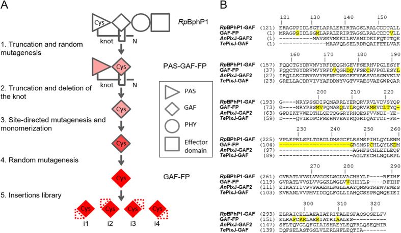
Figure 1. Rational design and molecular evolution of GAF-FP.
(A) Schematic representation of the directed molecular evolution of natural RpBphP1 resulted in GAF-FP and its small peptide insertion variants. The red color intensity depicts the fluorescence levels of the respective proteins. (B) Alignment of the amino acid sequence of GAF-FP with GAF domains of parental RpBphP1 and AnPixJ and TePixJ cyanobacteriochromes. The GAF-FP residues that differ from those of RpBphP1 are highlighted in yellow. Amino acid numbering follows that of RpBphP1.

