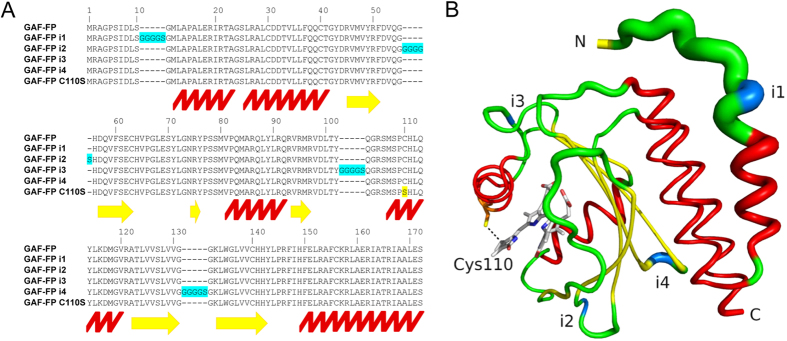
Figure 4. Design of insertion variants of GAF-FP.
(A) Alignment of the amino acid sequences of GAF-FP with GAF-FP i1, GAF-FP i2, GAF-FP i3, GAF-FP i4 and GAF-FP Cys110Ser templates. Insertions in GAF-FP are highlighted in blue, and Cys110Ser mutation is highlighted in yellow. Positions of α-helices are marked with the red zig-zag line, and positions of β-sheets are marked with the yellow arrows. Amino acid numbering follows that of GAF-FP without inserts. (B) Structural organization of GAF-FP based on the structure of GAF domain of parental RpBphP123. The thickness of the line indicates the magnitude of the structural β-factor. The α-helices colored in red, β-sheets are colored in yellow, and positions of the inserts are colored in blue.

