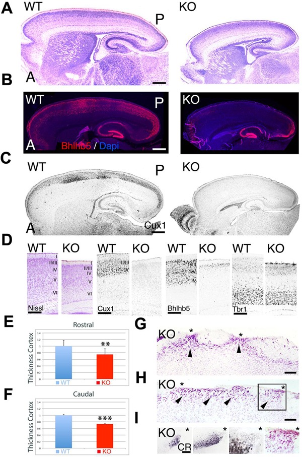
FIGURE 1.

Neuroanatomical comparison between wild-type (WT) and KO at P7. (A–C) Sagittal sections of WT and KO stained with Nissl (A) or immulabeled for Bhlhb5 (B) and Cux1 (C). Upper layer (UL) markers Bhlhb5 and Cux1 are reduced in the cortex of the KO. In (B), DAPI is shown as counterstaining. (D) Comparative profile between WT and KO cortical sections stained with Nissl or immunostained for the cortical layer markers Cux1 (II–IV), Bhlhb5 (II–V), and Tbr1 (VI). Cortical lamination is preserved in the KO but ULs are diminished and do not express the mature markers profile characteristic of UL neurons. (E,F) Quantitative analysis showing a significant reduction in cortical thickness by 25% at rostral levels (E, 0.75 ± 0.061∗∗, p = 0.0113, N = 8) and by 26% at caudal levels (F, 0.74 ± 0.009, p = 0.0001∗∗∗, N = 8) in the KO compared to WT. (G–I) In the KO, cortical ectopias (arrowheads) are observed in layers II/III and I using Nissl staining (G,H) and Calretinin (CR, I). Asterisks indicate cortical ectopias reaching the pial surface. A: anterior; I–VI: cortical layers 1–6; P; posterior. Scale bars: (A–C) (0.2 mm), (D) (125 μm), (G,H) (75 μm) and (I) (50 μm).
