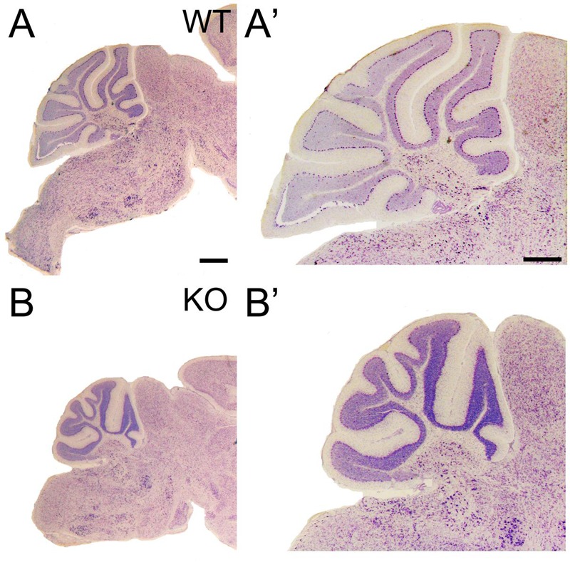
FIGURE 10.

Cerebellar volume is reduced and gyri foliation is poorly developed in the KO. (A–B’) P21 cerebellar sections of WT (A,A’) and KO (B,B’) stained with Nissl. (A’,B’) are high magnifications of (A,B), respectively. The volume of the cerebellum is reduced in KO compared to WT. It is noticeable the poorly developed gyri in some of the cerebellar lobes. Scale bars (A,B) (0.2 mm) and (A’,B’) (500 μm).
