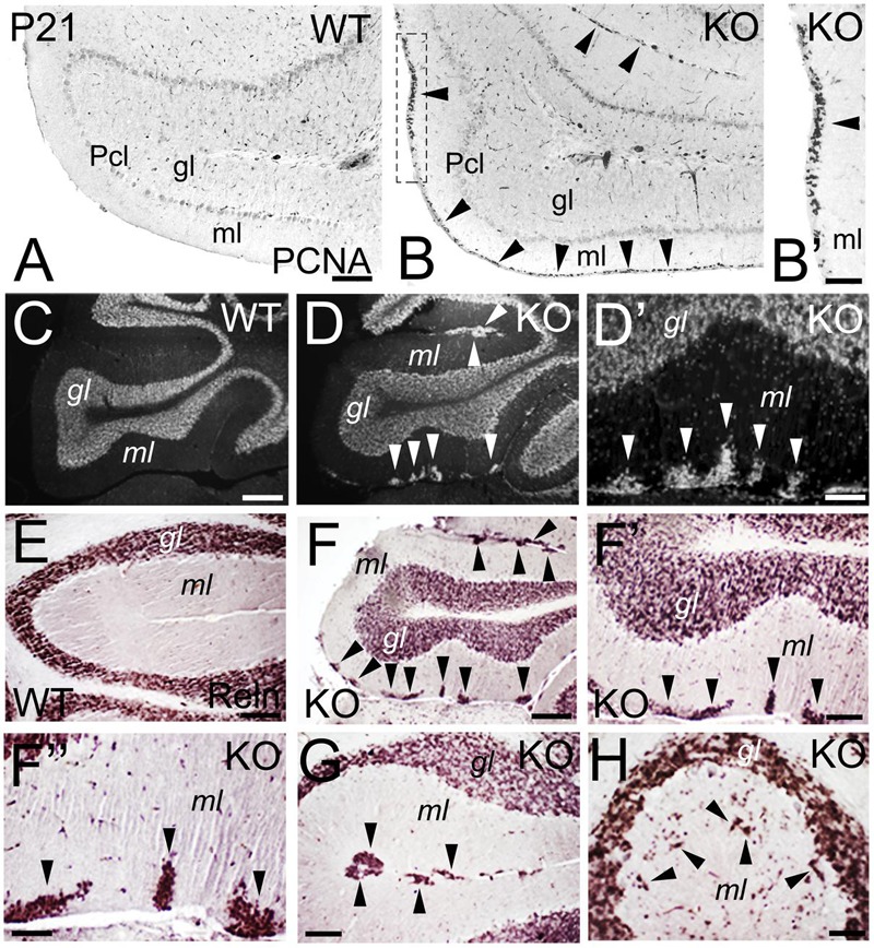
FIGURE 11.

Deficits in the migration and maturation of cerebellar excitatory granule cells suggest an ErbB4 dependent role. (A–B’) WT (A) and KO (B,B’) cerebellar sections at P21. A stream of proliferative PCNA+ granule cells (arrowheads) is observed in the molecular layer (ml) of the KO. In WT, no PCNA+ proliferation is observed because the cerebellum is already mature. (B’) high power view of (B). (C–H) WT (C,E) and KO (D,D’, F–H) cerebellar sections in adult mice. (C–D’) DAPI sections converted into monochromatic images showing granule cells in the granular layer (gl) of both WT and KO, in addition to ectopic granule cells in the molecular layer in the KO (arrowheads). (D’) high power view of (D). (E–H) Reelin (Reln)+ granule cells in WT (E) and KO (F–H) labeling the granular layer of both WT and KO and the ectopic granule cells observed only in the KO (arrowheads). (F’,F”) are high magnifications of (F). Pcl, Purkinje cell layer. Scale bars: (A,B) (100 μm), (B’) (50 μm), (C,D) (200 μm), (D’) (25 μm), (E) (50 μm), (F) (150 μm), (F’) (50 μm), (F”) (25 μm), and (G,H) (50 μm).
