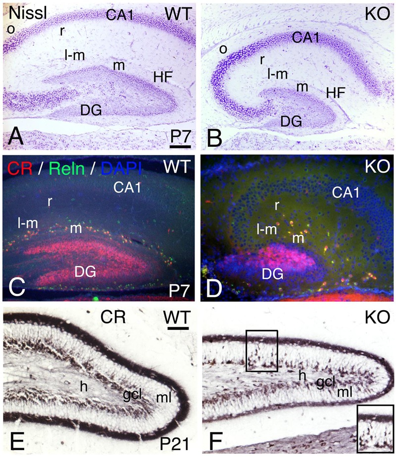
FIGURE 15.

Hippocampal development is compromised in the KO mice. (A,D) (P7) and (E,F) (P21) hippocampal sections of WT (A,C,E) and KO (B,D,F). (A,B) P7 Nissl staining shows a poorly developed hippocampus in the KO with a very rudimentary dentate gyrus (DG) and Cornus Ammonis (CA) fields. (C,D) At P7, the Calretinin (CR) and Reelin (Reln) interneuronal populations in the hippocampal strata lacunosum-moleculare (l-m), moleculare (m) and radiatum (r) are decreased in the KO compared to WT. (E,F) At P21, using Calretinin as a marker, I observe a fully developed dentate gyrus in WT. However, in the KO both dentate gyrus blades are thinner with a persistent migration of CR+ immature neuronal progenitors in the molecular layer (ml, inset in F). gcl, granular cell layer; h, hilus; HF, hippocampal fissure; o, strata oriens. Scale bars (A–D) (250 m) and (E,F) (125 μm).
