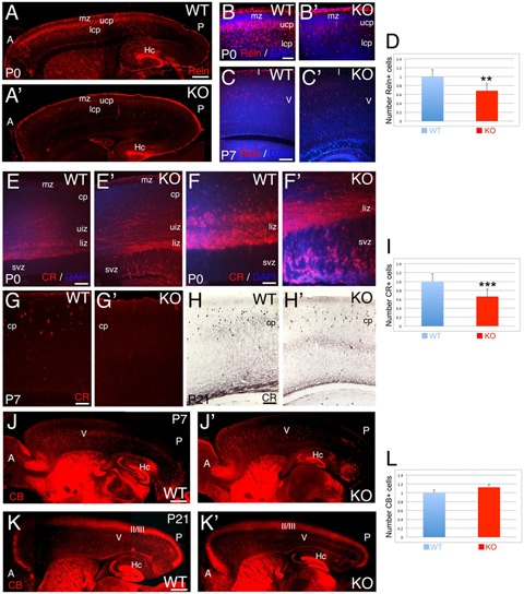
FIGURE 6.

Selective deficit of GABAergic interneuron subpopulations are observed in KO. (A–C’) Reelin (Reln)+ interneurons at P0 (A–B’) and P7 (C,C’) in WT (A,B,C) and KO (A’,B’,C’). At P0 WT, Reelin+ interneurons are expressed through a high-anterior to low-posterior gradient at the interface between mz and upper cortical plate (ucp) with radially oriented interneurons also present in the lower cortical plate (lcp). In the KO, the Reelin+ interneuronal gradient is very rudimentary with a severe reduction in the number of Reelin+ interneurons. (B,B’) are high power views of (A,A’), respectively. At P7 WT, Reelin+ interneurons are mostly located in layer V in the cortical plate (cp), whereas in KO, the density of Reelin+ interneurons is reduced and they are more dispersed throughout the cortical plate. DAPI is shown as counterstaining. (D) At P21, Reelin+ interneurons are significantly reduced by 32% in the cortex of KO compared to WT (0.68 ± 0.07, p = 0.0261∗∗, N = 4). (E–H’) Calretinin (CR)+ interneurons at P0 (E–F’), P7 (G,G’), and P21 (H,H’) in WT (E,F,G,H) and KO (E’,F’,G’,H’). At P0, WT CR+ interneurons are radially oriented in the lower intermediate zone (liz) with subsets of CR+ interneurons already migrating in the upper intermediate zone (uiz). In the KO, a dense plexus of CR+ fibers and tangentially oriented CR+ interneurons are observed in the upper and lower intermediate zones, where the vast majority of radially oriented CR+ interneurons are located in the svz. (F,F’) are high power views of (E,E’), respectively. At P7, CR+ interneurons are located in the upper cortical plate in WT and absent in the KO. DAPI is shown as counterstaining. At P21, CR+ interneurons are densely observed in the cortical plate in WT, with a severe reduction in the KO. (I) At P21, a statistically significant reduction of CR+ interneurons by 35% is observed in the KO compared to WT (0.66 ± 0.07, p = 0.0004∗∗∗, N = 9). (J–K’) Calbindin (CB)+ interneurons at P7 (J,J’) and P21 (K,K’) in WT (J,K) and KO (J’,K’). At P7, CB+ interneurons are mostly located in layer V in both WT and KO, but at posterior levels their number is severely reduced with defects in their distribution pattern. At P21, the majority of CB+ interneurons are located in layers II/III and V. DAPI is shown as counterstaining. L: Quantification of CB+ interneurons indicates no statistically difference between WT and KO at P21 (1.12 ± 0.01, p = 0.186, N = 8). A, anterior axis; Hc, hippocampus; I, cortical layer 1; II/III: cortical layers 2/3; P, posterior axis; V, cortical layer 5. Scale bars: (A,A’) (0.2 mm), (B,B’) (50 μm), (C,C’) (100 μm), (E,E’) (100 μm), (F,F’) (75 μm), (G–H’) (100 μm), and (J–K’) (0.2 mm).
