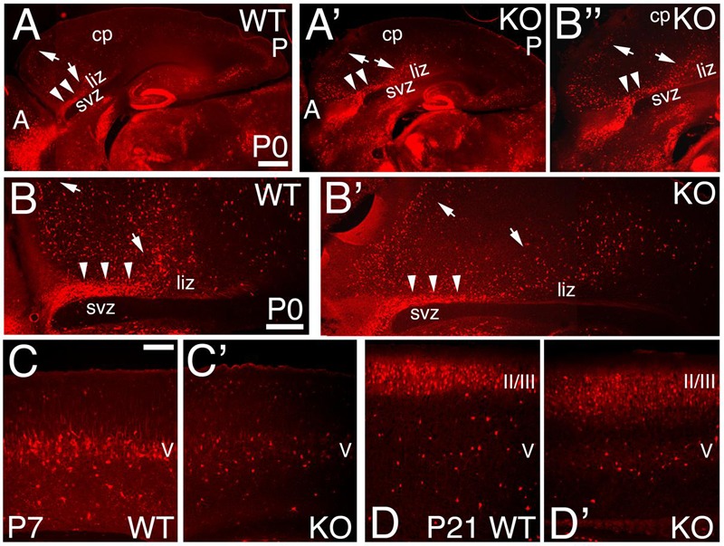
FIGURE 8.

Expression pattern of Calbindin positive interneurons in the KO mice. (A–D’) Sagittal sections at P0 (A–B”), P7 (C,C’), and P21 (D,D’) in WT (A,B,C,D) and KO (A’,B’,B”,C’,D’) labeled with Calbindin (CB). At P0, CB+ interneurons reach the cortex using the interphase (arrowheads) between the lower intermediate zone (liz) and svz; from which they migrate radially to their final cortical position (arrows) without entering the SVZ. This pattern is preserved in the KO. (B,B”) are high magnifications of (A,A’), respectively. By P7, CB+ interneurons are mostly located in layer V in WT, but they are severely reduced in the KO. At P21 WT, CB+ interneurons are densely packed in layers II/III and spread throughout the deep layers. In KO, CB+ interneurons are spread throughout layers II/III and localized in layer V. cp, cortical plate; II/III and V, cortical layers 2/3 and 5. Scale bars, (A,A’) (0.2 mm), (B,B’) (500 μm), (B”) (0.2 mm) and (C–D’) (100 μm).
