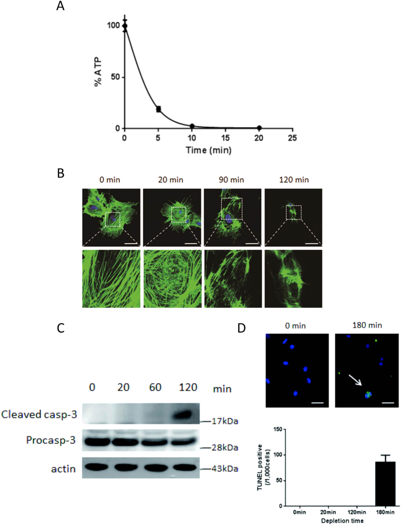
Figure 1. ATP depletion disrupts actin cytoskeleton and induces apoptosis.
Incubation of cultured mouse differentiated podocytes with 100 mM 2-deoxyglucose (2-DG) and 10 μM antimycin rapidly depleted intracellular ATP within 15 min (A). ATP depletion induced a granular pattern of phalloidin-labeled actin at 20 min compared with actin stress fibers under normal conditions. Subsequently, actin distribution became markedly deranged, with loss of stress fibers at 90 min, and complete disruption at 120 min (B). Cleaved caspase 3, an activation marker of caspase 3, was evident at 120 min in the cropped gel (C) followed by the appearance of terminal deoxynucleotidyl transferase-mediated dUTP-biotin nick end-labeling (TUNEL) positive cells (arrow) at 180 min (D). All blots in the each photo were run under the same experimental conditions. Size markers indicate the cropped level in each figure. Full length gels and blots for each figure are shown in Supplemental Figure 2. Bar, 50 μm.

