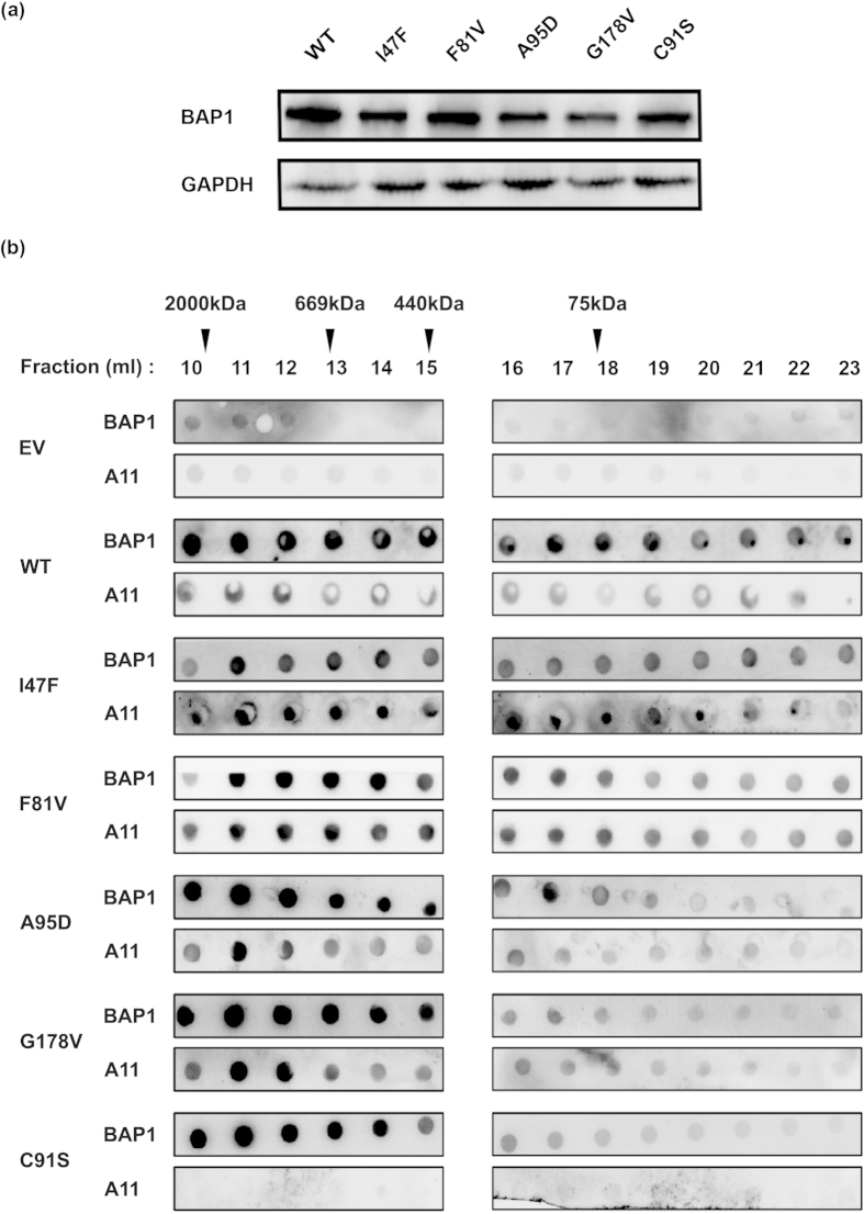
Figure 3. Characterization of BAP1 amyloid aggregates by dot blot assay.
(a) HEK293T cells were transfected with Flag-HA wild type (WT), empty vector (EV) and mutant (I47F, F81V, A95D, G178V and C91S) plasmids. After 48 h of transfection, cells were harvested and immunoblot analysis was performed. GAPDH was used as a loading control. Similar expression level of wild type and mutant BAP1was detected. (b) Dot blot analysis of cellular extracts of BAP1 wild type (WT) and its mutants (I47F, F81V, A95D, G178V and C91S) was performed. Chromatographic fractions after gel filtration were dot blotted and incubated with anti-BAP1 and anti-A11 antibodies. Reactivity of A11 and BAP1 were normalized to total protein content. Molecular weight and fraction numbers are indicated in the upper panel.

