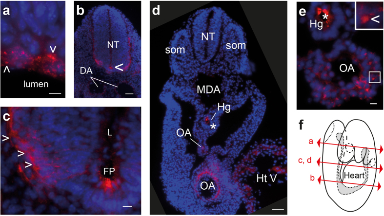
Figure 1. PrPC expression pattern in the nervous and cardiovascular systems from early developing mouse embryos.
PrPC (red) and nuclear marker 4’,6-diamidino-2-phenylindole (DAPI, blue) staining of transverse sections from FVB/N mouse embryos at E9 in the developing nervous (a–c) and cardiovascular systems (d,e). Section plans are indicated (f). PrPC was observed at the level of the apical face of the neuroepithelium in the head region (presumptive telencephalon; a), in the neural tube (NT) in the trunk region (b), and particularly in the floor plate (FP, c). The arrowhead indicates PrPC-positive differentiating cells in the mantle zone (c). Low-exposure acquisition of two juxtaposed embryo sections from the proximal and caudal trunk regions showed strong PrPC expression in the omphalomesenteric artery (OA) and in the embryonic heart ventricle (HtV) (d). Higher magnification of the omphalomesenteric artery (OA) (e). The asterisks indicate artefact signals found in FVB/N and in Prnp−/− mice (supplemental Figure S1), likely due to nonspecific Fc binding to maternal blood, and the arrowheads highlight specific punctuate or patchy PrPC signals. Scale bar: 10 μm, except in (b,d) 50 μm. DA: dorsal aorta, Hg: hindgut, L: lumen, MDA: midline dorsal aorta, som: somite.

