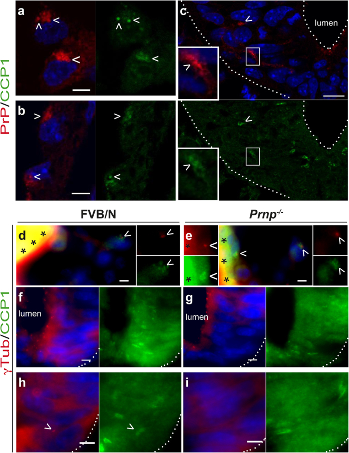
Figure 7. Detection of cytosolic carboxypeptidase 1 in the mouse embryo.
Transverse sections from FVB/N (a–d,f,h) and Prnp−/− (e,g,i) E9 mouse embryos co-stained for CCP1 (green), DAPI (blue) and PrPC (a–c) or γTub (d–i) (red) are shown (representative images of n = 4 embryos analysed). The dotted lines and asterisks indicate the boundaries of the neural tube and artefact signals, respectively. PrPC and CCP1 co-localization (arrowheads) in the yolk sac (a), omphalomesenteric artery (b) and the mantle zone of the neural tube (c, inset), as observed by confocal microscopy. Note that no co-localization is observed at the apical face of the floor plate (c). γTub and CCP1 co-staining in the yolk sac at the base of the primary cilium (arrowheads; d,e). Expression patterns of γTub and CCP1 in the neural tube (f–i). Juxtaposition is rarely observed (arrowheads in h). Scale bar: 5 μm, except in c: 10 μm.

