
Figure 1.
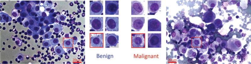
Sample mesothelial nuclei are show in this figure. The left-most image is taken from a benign effusion, and the rightmost image is taken from a mesothelioma patient. Sample selected nuclei from each type are given in the center, showing the similarity of different types of nuclei. [Color figure can be viewed in the online issue, which is available at wileyonlinelibrary.com.]