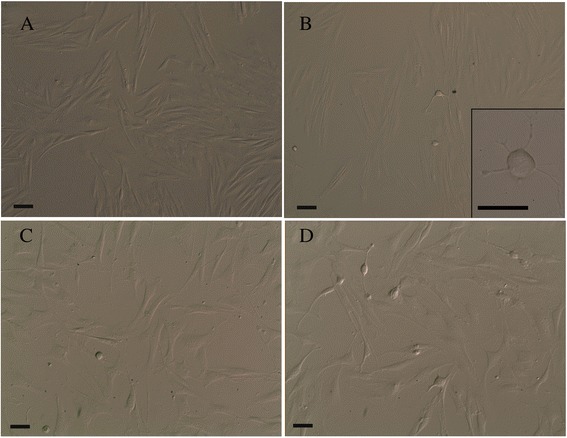
Fig. 1.

Phase contrast images of cultured EC-MSCs. Homogeneous fibroblast-like cells (spindle-shaped in appearance) at P0 (a); among fibroblast-like cells, some adherent cells with a different shape (spherical or oval) and size were distinguishable at P2. Insert: a spherical cell body with thin cytoplasmic extensions (b). Spherical cells without processes were free floating in the medium, whereas cells with neuronal- and glial-like morphology with extending processes were firmly adherent to the bottom of the flasks at P3 (c). Cells also showed unipolar, bipolar or multipolar extensions at P3 (d). Bars, 50 μm. EC-MSCs equine cadaver mesenchymal stem cells
