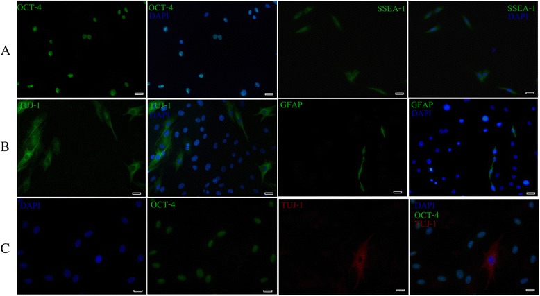
Fig. 4.

Immunocytochemistry analysis of pluripotency (OCT-4 and SSEA-1), neuronal (TUJ-1) and glial (GFAP) markers in cultured EC-MSCs at P3. High percentage of cells expressed OCT-4 and SSEA-1 (OCT-4, green; SSEA-1, green; DAPI, blue) (a). A fibrillar cytoplasmic staining was detected with TUJ-1 while the GFAP expression was seen in both the cytoplasm and the cytoplasmic extensions of only a few cells (TUJ-1, green; GFAP, green; DAPI, blue) (b). Analysis of TUJ-1 and OCT-4 localization showed that TUJ-1-positive cells exhibited a neuronal-like appearance and were OCT-4 negative (TUJ-1, red; OCT-4, green; DAPI, blue) (c). Bars: 20 μm. OCT-4 pluripotent transcription factor, SSEA-1 stage-specific embryonic antigen-1, TUJ-1 neuron-specific class III beta-tubulin, GFAP glial fibrillary acidic protein, EC-MSCs equine cadaver mesenchymal stem cells, DAPI 4',6-diamidino-2-phenylindole
