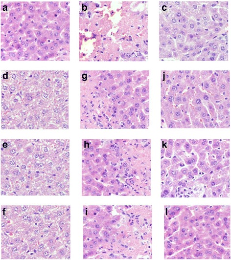
Fig. 4.

Representative photomicrographs of liver histopathology (400×). a: liver in mice of NC groups showing normal cellular architecture of hepatic tissue; (b): liver of mice in MC groups after injection of STZ (120 mg/Kg) showing cellular degeneration, hepatocyte necrosis, and lipid droplet accumulation; (c): liver of mice in PC groups; (d-f): liver of mice fed with Ac-, Al-, and En-MZPS at dosage of 800 mg/Kg showing mild architectural damage; (h-i): liver of mice fed with Ac-, Al-, and En-MZPS at dosage of 400 mg/Kg showing mild architectural damage with few showing abnormal structure; (j-l): liver of mice fed with Ac-, Al-, and En-MZPS at dosage of 200 mg/Kg showing almost normal histology similar to control mice
