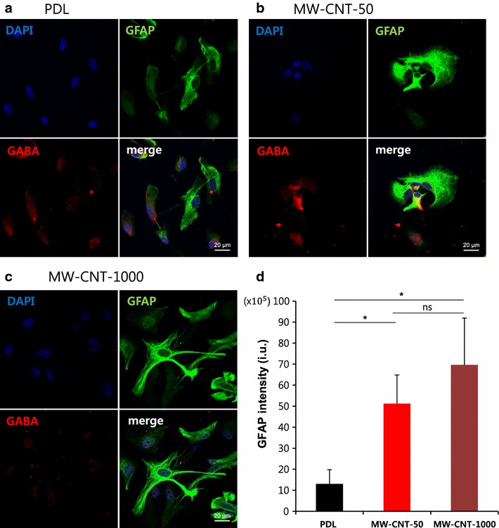
Fig. 4.

Astrocytic GABA on MW-CNTs spreads into cell processes. a–c Immunostaining of GABA using anti-GABA (red), anti-GFAP (green) antibody in primary cortical astrocytes on MW-CNT and PDL coverslips. a shows distribution of astrocytic GABA near the nucleus. b shows astrocytes on MW-CNT-50 to have a rounder shape compared to those on PDL and c shows distinct cell–cell interactions on MW-CNT-1000. a–c shows that intracellular GABA on MW-CNT has spread into cell processes (b, c) compared to the PDL coverslip (a); Scale bar 20 μm. d Graph shows MW-CNTs increase GFAP immunoreactivity compared to PDL (*p < 0.05); PDL (n = 7), MW-CNT-50 (n = 7), MW-CNT-1000 (n = 7). Fluorescence intensity (i.u.) = cell intensity − (area × mean fluorescence of background)
