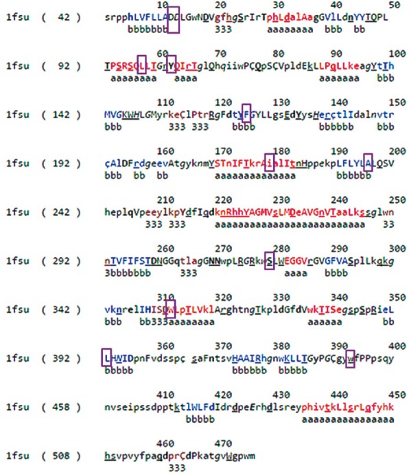
Fig. 1.

ARSB sequence and structural analysis: Protein Data Bank (PDB) file output of JOY for protein P15848. Residues form alpha helices, beta strands and 310 helices are coloured red, blue and maroon, respectively. Solvent accessible and inaccessible residues are written in lower and upper cases, respectively. Residues that bind to the main-chain amide using hydrogen bonds are written in bold and the ones that bind to main-chain carbonyl are underlined. Residues with positive phi torsion angles are italicised and the ones that form disulphide bond have a cedilla. Positions of mutated residues have been enclosed in a purple box.
