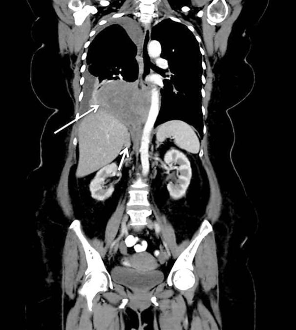
Figure 1:

Radiological features of β-hCG secreting lung adenocarcinoma. Coronal reconstruction contrast enhanced computed topography (CT) abdomen and thorax shows a heterogenously enhancing right lower lobe mass (long arrow) which displaces and abuts the descending thoracic aorta. The right adrenal is enlarged in keeping with adrenal metastasis (short arrow).
