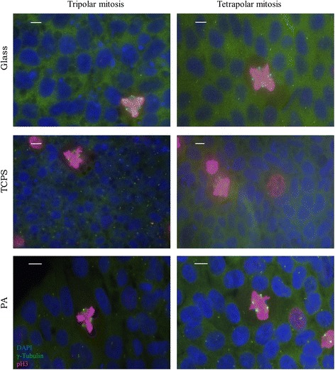
Fig. 2.

Commonly observed multipolar mitoses in 19-9-11 iPSCs cultured on substrates of varied stiffness. Nuclei are labeled in blue (DAPI), γ-tubulin is labeled in green while pH3 is labeled in red. The rows indicate the substrates human iPSCs were cultured on. Tripolar mitoses are characterized by 3 spindle poles (green γ-tubulin foci). Tetrapolar mitoses are characterized by 4 spindle poles. These types of abnormalities are included in the percentage of abnormal mitoses calculated in Figs. 3 and 4. Scale bars: 10 μm
