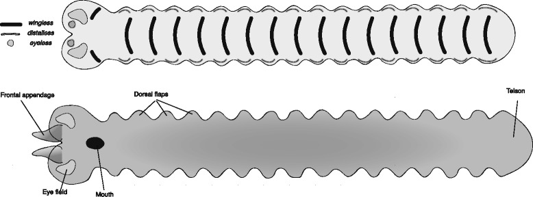
Fig. 3.

Reconstructed germband of Opabinia regalis. The upper panel represents an early stage germband with the expression of select marker genes mapped onto it. The lower panel represents a late stage germband, annotated with the adult fate of the main embryonic features. Note the close antero-medial position of the paired frontal appendage anlagen. These are assumed to fuse at dorsal closure to give a single appendage. The origin of the eyes has not been reconstructed in detail, and a broad “eye field” is shown instead
