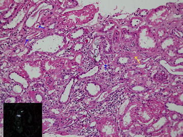
Fig. 2.

Histology (haematoxylin and eosin stain) of renal biopsy of case 2 showing acute on chronic oxalate induced tubulo-interstitial nephritis. One tubule contains oxalate crystals (black arrow) which polarize with polarizing light (inset). Evidence of chronic interstitial nephritis, i.e. intersititial fibrosis (yellow arrow) and tubular atrophy (blue arrow) are present. The interstitium contains a moderate numbers of lymphocytes
