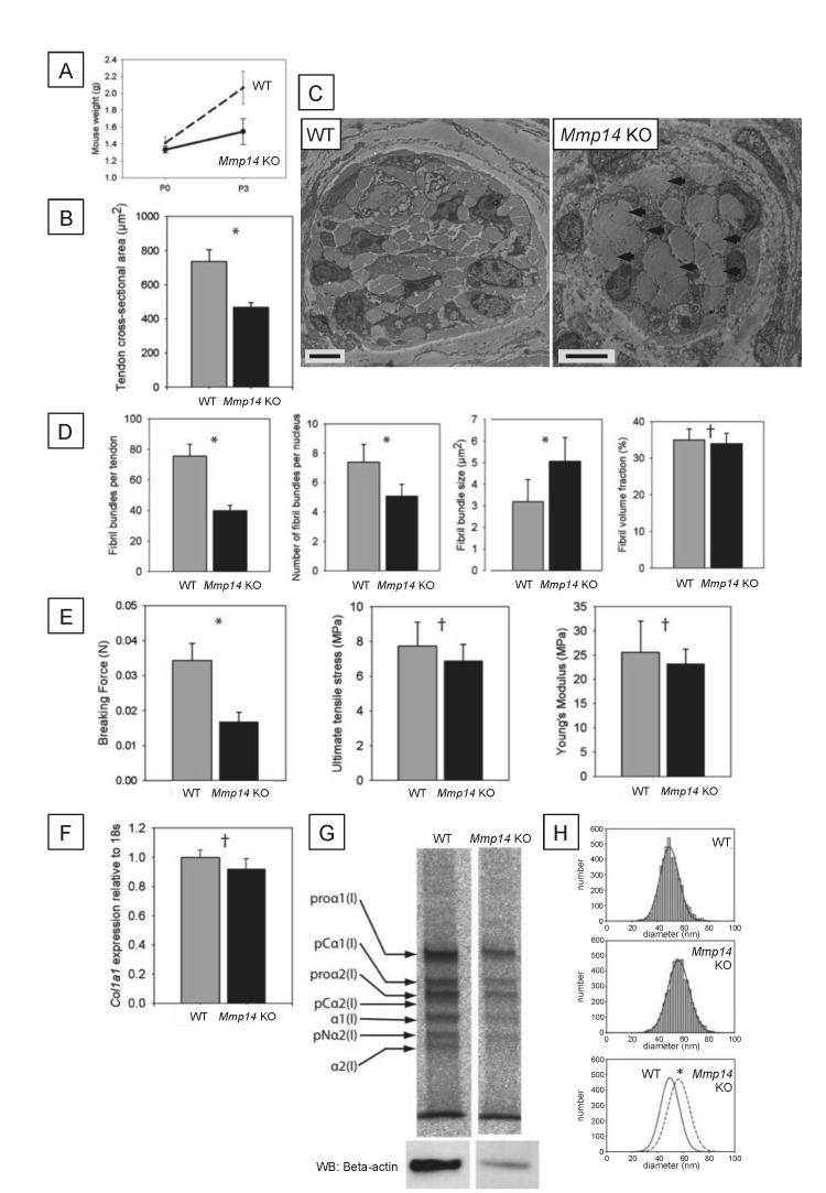
Figure 1. Neonatal Mmp14-deficient mice have small and weak tendons.
(A) Weight of wild type (WT) and Mmp14 knockout (Mmp14 KO) littermates at birth (P0) and at 3 days postnatal (P3). (B) The cross-sectional transverse area of P0 Mmp14 KO tendons is significantly smaller than WT tendons. (C) TEM images of P0 tail tendon demonstrate that KO tendons are smaller and show dysmorphic, enlarged bundles of collagen fibrils (arrowhead). Scale bars 5 μm. (D) KO tendons have fewer, larger fibril bundles, but the FVF is not different to WT tendons. (E) KO tendons are weaker than WT tendons but have normal mechanical properties after adjusting for differences in size. (F) Analysis of Col1a1 mRNA by qPCR in P0 tendons revealed no difference in gene expression. (G) 14C-proline labeling of collagen demonstrated normal collagen processing at P7 in WT and KO tail tendon. (H) Fibril diameter distributions of KO and WT tail tendon at P0 revealed significantly increased fibril diameters in KO mice. Bars show SEM. *p < 0.05, †p > 0.05 (t-tests).


