. 2015 Sep 21;4:e09345. doi: 10.7554/eLife.09345

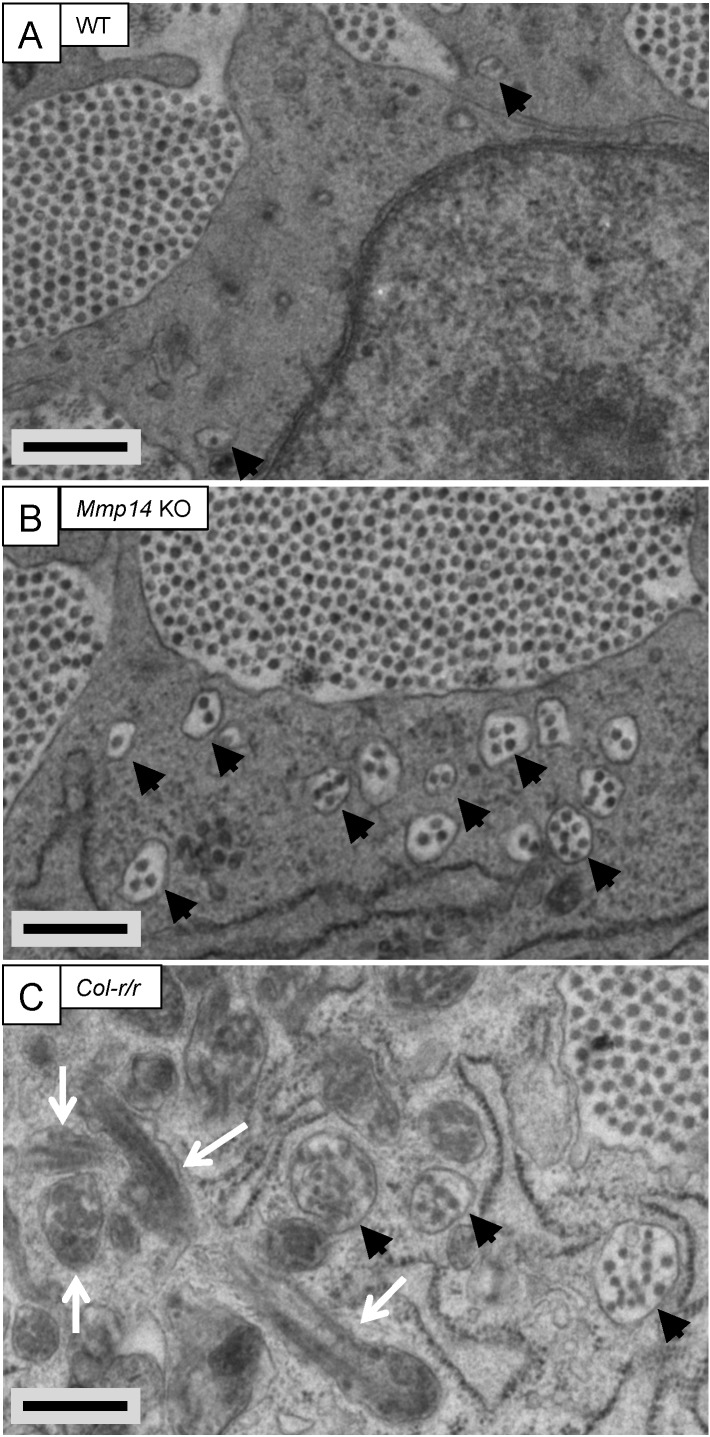
Figure 2. Fibricarrier analysis of wild-type, Mmp14 KO, and Col-r/r embryonic tail tendon.

Tail tendons at E15.5 of development from (A) wild-type, (B) Mmp14 KO, and (C) Col-r/r mice. Black arrowhead, recessed fibripositor (electron lucent)-containing collagen fibrils. White arrow, enclosed electron-dense compartment. Scale bars 500 nm.

DOI: http://dx.doi.org/10.7554/eLife.09345.005

Figure 2.

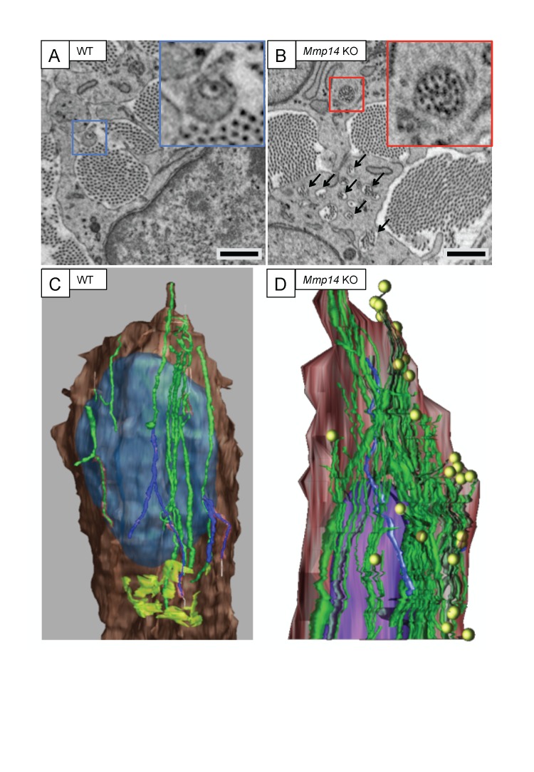
Figure 2—figure supplement 1. Mmp14-deficient mice have prominent recessed fibripositors.

Figure 2—figure supplement 1.

SBF-SEM of (A) WT and (B) Mmp14 KO tendons at E15.5 showing fibripositors in WT tendons (blue box) and intracellular collagen fibril-containing compartments in Mmp14 KO tendons (red box; arrowheads). Scale bars 1 µm. 3D reconstruction of SBF-SEM data from (C) WT and (D) Mmp14 KO tendon cells. Brown, cytoplasm. Purple/blue, nucleus. Green, internal collagen fibril compartments. Yellow circles, fibril ends. Lime green (in WT only), Golgi apparatus.
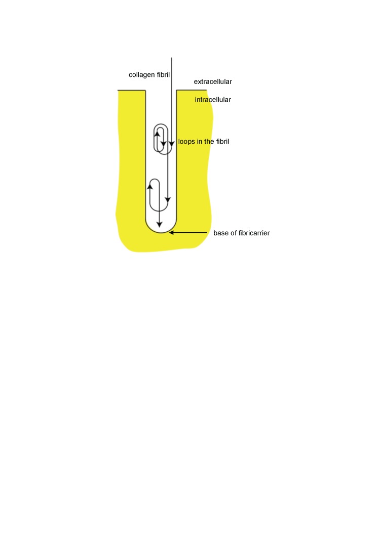
Figure 2—figure supplement 2. Schematic showing looping of collagen fibrils in recessed fibripositors.

Figure 2—figure supplement 2.

Diagrammatic representation of a recessed fibripositor with looping of a collagen fibril. Not drawn to scale.