. 2015 Sep 21;4:e09345. doi: 10.7554/eLife.09345

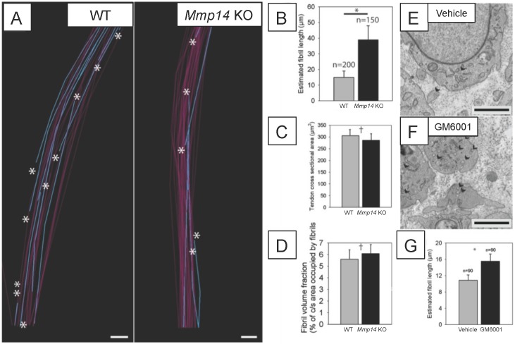
Figure 3. Deficiency in MMP14 activity results in fewer collagen fibrils.

(A) 10 µm-deep (z-axis) slices of 3D reconstructions of SBF-SEM data taken from of WT and Mmp14 KO embryonic tendons at E15.5 showing collagen fibrils (blue) with a tip (marked by asterisks) found within the volume. Purple fibrils passed through the volume and so did not have tips in the reconstruction. Scale bars 500 nm. (B) Quantification of mean fibril length based on the number of tips identified shows that E15.5 WT fibrils are shorter than fibrils in age- and anatomical position-matched tail tendons from KO mice (308 and 266 fibrils tracked, respectively). (C) Tendon cross-sectional area and (D) FVF are not different at E15.5 KO tendons. (E, F) Electron microscopy of tendon-like constructs cultured in the presence of MMP inhibitor GM6001 (10 µM in 0.1% DMSO) show increased number of recessed fibripositors (arrowheads) compared to vehicle control. Scale bars 1 µm. (G) Increase in calculated mean fibril length in GM6001-treated tendon-like constructs. Bars show SEM. *p < 0.05, p > 0.05 (t-tests).

DOI: http://dx.doi.org/10.7554/eLife.09345.010

Figure 3.

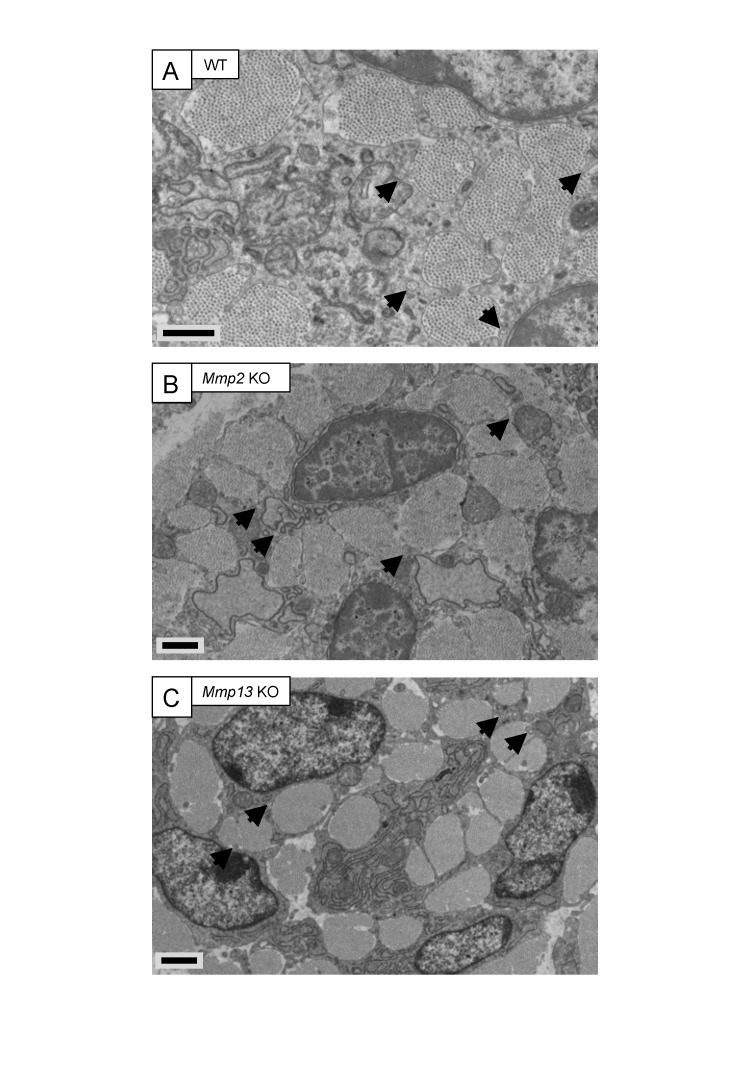
Figure 3—figure supplement 1. Embryonic tendons deficient in Mmp2 or Mmp13 do not have overt tendon phenotypes.

Figure 3—figure supplement 1.

Electron microscopy images of embryonic tendons from (A) WT, (B) Mmp2 KO, and (C) Mmp13 KO mice. Arrowheads indicate the locations of fibripositors. Scale bars 1 µm.
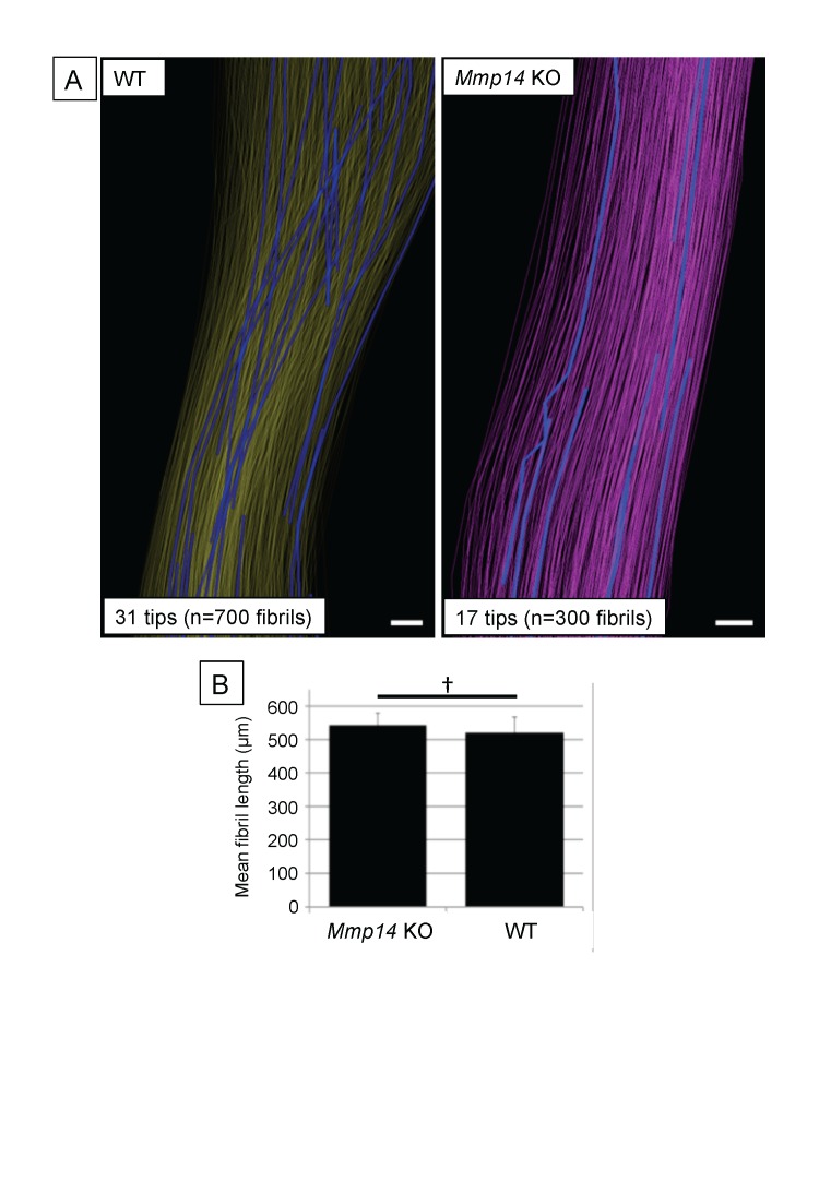
Figure 3—figure supplement 2. Deficiency in MMP14 activity results in fewer collagen fibrils tips at P0.

Figure 3—figure supplement 2.

(A) 3D reconstruction from SBF-SEM analysis of P0 WT and Mmp14 KO tendons. Scale bars 2 µm. (B) Estimated mean fibril length 645 ± 14 µm WT and 674 ± 24 µm Mmp14 KO shows that the absence of MMP14 does not affect fibril length at birth. Bars show SEM. p > 0.05 (t-test).
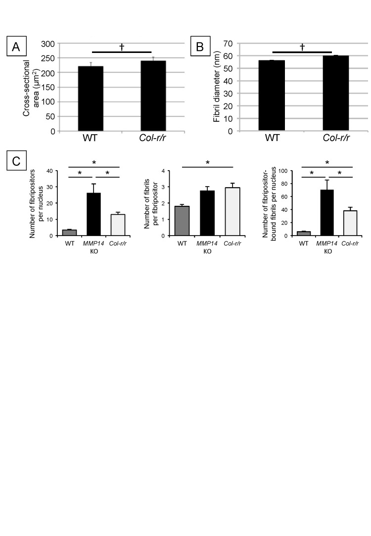
Figure 3—figure supplement 3. Quantitative analysis of Col-r/r embryonic tendons.

Figure 3—figure supplement 3.

Measurement of tendon cross-sectional area (A) and collagen fibril diameter (B) shows no significant difference between WT and Col-r/r mice at E15.5. p > 0.05 (t-tests). (C) Quantitation of fibril-containing carriers in WT, Mmp14 KO, and Col-r/r embryonic tenocytes. *p < 0.05 (one-way ANOVA). Bars show SEM.