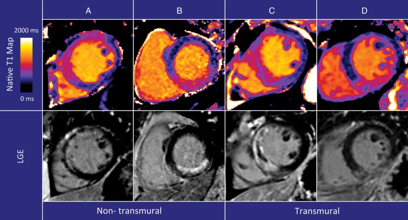
Figure 3.
Native T1 maps in non-transmural and transmural infarction. Native T1 maps (top row) and LGE of the infarction (bottom row). Both in patients with non-transmural and transmural infarctions, the abnormality in native T1 abnormality appears transmural. The culprit lesion was the left anterior descending artery in images in (A and C) and the right coronary artery in (B and D).

