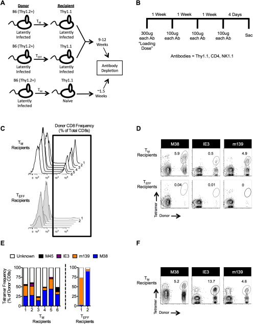
Figure 7. TM cells persist in latently-infected, immune replete mice and expand when host immunity is lost.
(A) Schematic of experimental design. Age matched B6 and Thy1.1 mice were infected with 1×10⁶ pfu MCMV-Smith. Following the establishment of viral latency (>8 weeks post-infection), either TM or TEFF cells from the B6 donors were transferred, as described in the Methods, into the latently-infected Thy1.1 recipients or into naïve Thy1.1 mice. Latently-infected recipients were rested for 9-12 weeks, while the naïve recipients were rested for approximately 1.5 weeks. (B) Antibody depletion schedule. (C-E) The presence of tetramerpos donors was analyzed by flow cytometry immediately following the depletion schedule. Data were collected from two independent experiments (n = 6 total). Three mice from each group were depleted 9 weeks after the transfer; three mice from each group were depleted 12 weeks after transfer. (C) Histograms of donor T cells within each individual recipient. (D) Representative FACS plots of tetramerpos donors immediately following the depletion regimen. (E) Frequency within each individual recipient of each analyzed tetramer as a percent of total donor CD8+ cells. TEFF recipients 3-6 are excluded because they did not have a donor population. (F) Tetramer staining was performed 11 weeks after depletion in one experiment described above (n = 3).

