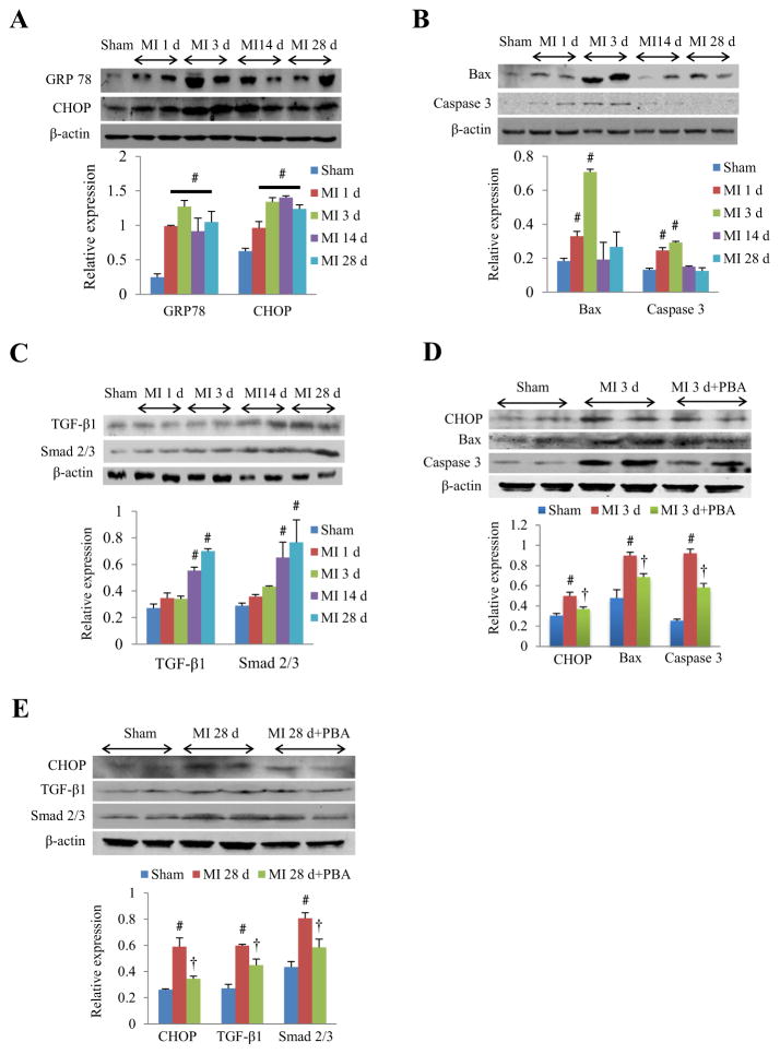
Fig. 1.
ER stress response in cardiac apoptosis and fibrosis after MI. (A) Western blots and quantitative analysis of ER stress response markers (GRP 78 and CHOP) in the whole heart homogenates of mice in the early and late phase of MI. (B) Western blots and quantitative analysis of pro-apoptotic proteins (Bax and caspase 3) in the whole heart homogenates of mice in the early and late phase of MI. (C) Western blots and quantitative analysis of pro-fibrotic proteins (TGF-β1 and Smad 2/3) in the whole heart homogenates of mice in the early and late phase of MI. (D) Representative Western blots and quantitative analysis of CHOP, Bax and caspase 3 from whole heart homogenates at 3rd day of MI with or without 4-PBA (20 mg/kg/d). (E) Representative Western blots and quantitative analysis of CHOP, TGF-β1 and Smad 2/3 from whole heart homogenates at 28th day of MI with or without 4-PBA (20 mg/kg/d). #P < 0.05 vs. sham; †P < 0.05 vs. MI; n = 4–6 in each group.

