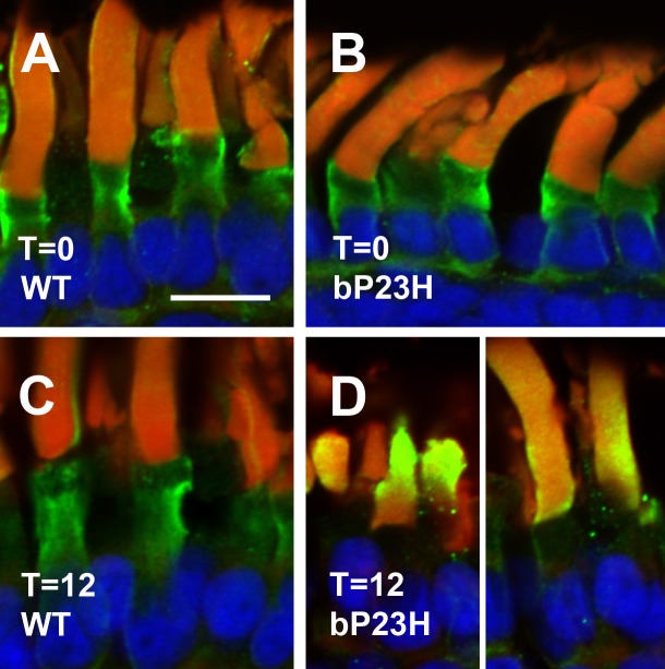
Figure 5.
Arrestin localization is abnormal in light-exposed bP23H retinas. (A) Dark-reared, WT. (B) Dark-reared, bP23H. (C) WT, 12 hours of light exposure. (D) bP23H, 12 hours of light exposure. Arrestin immunolabeling is shown in green, wheat germ agglutinin in red, and Hoechst 33342 in blue. Distribution of arrestin is altered in light-exposed bP23H photoreceptors, with increased outer segment labeling. Scale bar: 10 μm.

